Lattice defect mapping
Mapping the Invisible: How 3D Defect Imaging Reveals the Heart of a Gemstone
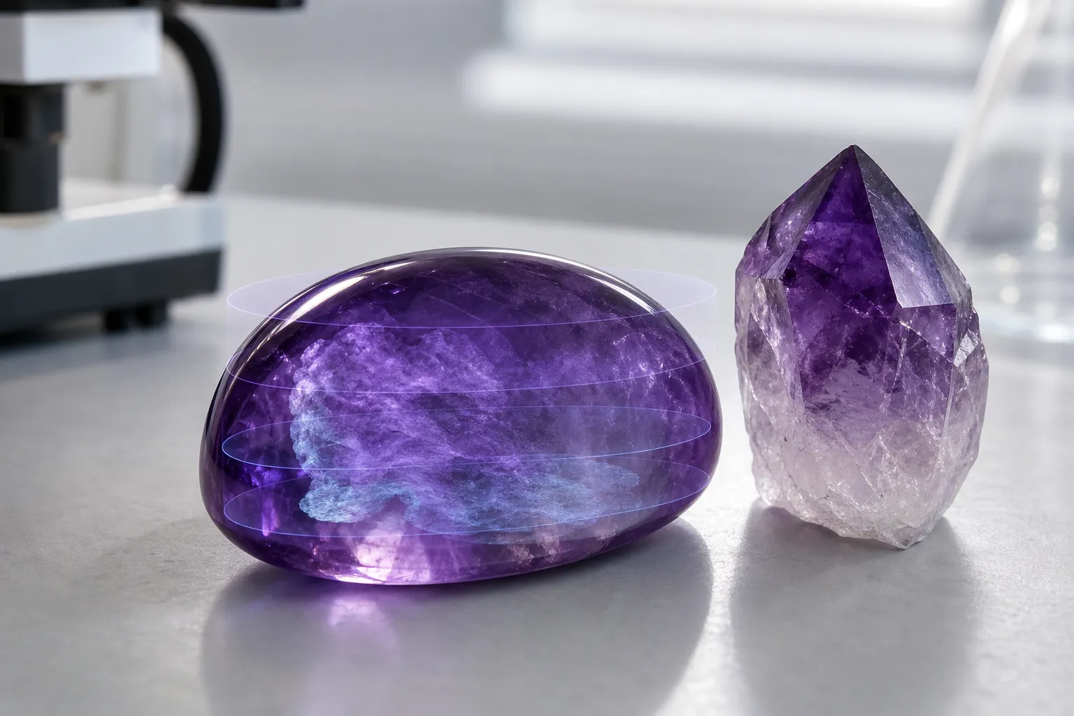
A polished amethyst can look quiet at the surface: violet zoning, glassy luster, perhaps a faint veil or fracture catching the light. Lattice defect mapping asks what the eye cannot settle from that surface alone. In plain terms, 3D defect imaging can help readers picture a gemstone as a volume, not just a face; it may show where internal signal changes, structural irregularities, or defect-related features are distributed through the stone.
That is useful, but it is not a verdict. A defect map may support a closer look at invisible gemstone features. By itself, it does not establish origin, market value, authenticity, treatment history, or spiritual meaning.
broader context
Broader amethyst guide
This narrower page lands better after the broader amethyst context page.
What 3D Lattice Defect Mapping Means
A gemstone is more than color. It is a mineral structure shaped by growth conditions, trace chemistry, internal strain, inclusions, fractures, and small irregularities in the crystal lattice. Some of those features can be seen under magnification. Others are too small, too faint, or too spatially complicated to read from the outside.
3D lattice defect mapping is best understood as a way of locating and comparing internal signals across a gemstone volume. Instead of treating one measurement point as the whole story, mapping asks where a signal appears, how strongly it appears, and whether it changes from one region to another. For a gemstone reader, the key idea is distribution.
That matters for amethyst because color zoning already trains the eye to think spatially. A crystal may be pale near one end and deeper violet near another; a cluster may preserve growth faces that hint at changing conditions. Defect distribution analysis extends that habit into features the unaided eye cannot resolve. It suggests that a stone’s interior may have a map-like character, even when the surface looks even.
The map still needs interpretation. Without qualified method details, comparison material, and source support, a 3D image should be treated as investigative context rather than a final judgment.
How Spectroscopy Can Become a Spatial Picture
Terms often connected with gemstone defect imaging include Raman spectroscopy, photoluminescence, and spatial distribution analysis. For this page, the point is not to turn the reader into an instrument specialist. The useful question is simpler: how can repeated measurements become a picture of internal variation?
Raman spectroscopy signals are commonly discussed as material-response signals. A single measurement may describe one point or a small region. A mapped study can collect many readings across an area or volume, then compare how the signal changes from place to place. When the method is well documented, that kind of mapping may help visualize internal differences instead of reducing the stone to one averaged reading.
Photoluminescence mapping follows a similar reader-facing idea. Signal behavior can vary across a material, and gemstones are not always internally uniform. Growth zones, strain patterns, defects, and inclusions may appear in some regions and not in others.
The careful takeaway is narrow: spectroscopy signals can be treated as points of information, and mapping can arrange those points into a spatial picture. The harder work is deciding what that picture means.
What Invisible Features It May Help Investigate
For a collector, “invisible” should not mean mystical. It simply means a feature is not readily seen in normal viewing, even if it may be studied with appropriate tools. Internal gemstone features can include variations in structure, signal behavior, growth zones, defects, inclusions, or stress-related patterns.
Questions a Map Can Help Ask
- Is the internal signal concentrated in one zone or spread through the crystal?
- Does a signal follow visible color zoning, or does it cross those zones differently?
- Are there regions that look internally distinct even when the surface appears continuous?
- Does the spatial defect distribution suggest a local feature rather than a whole-stone pattern?
- Is the mapped result consistent enough to invite further gemological review?
These are questions, not conclusions. A map of gemstone lattice defects may reveal complexity, but complexity alone does not equal rarity. It does not automatically separate natural from treated material. It does not turn a decorative specimen into a high-value collector piece. It also does not validate symbolic claims about what the crystal represents in a room, collection, or meditation practice.
For amethyst, that separation matters. A violet crystal may be chosen for beauty, geological interest, display presence, or spiritual symbolism. Defect mapping belongs to the material side of the conversation. It can refine how one thinks about the stone’s interior; it should not be used as a shortcut for meanings that belong to personal or cultural interpretation.
What Makes the Answer Stronger or Weaker
The phrase “3D lattice defect mapping” can sound precise, but the strength of any claim depends on conditions that are easy to miss.
Method
Raman spectroscopy, photoluminescence mapping, and other forms of materials characterization do not all measure the same response. A colorful internal image should not be treated as self-explanatory.
Sampling Pattern
A sparse set of measurement points cannot support the same reading as a dense, well-described spatial distribution. If only one face, one slice, or one small zone is examined, the result may not describe the whole gemstone.
Specimen
A faceted stone, rough crystal point, geode section, and cluster each present different practical problems. Surface geometry, inclusions, fractures, and color zoning may affect what can be examined and how confidently a result can be read.
Comparison Material
A mapped signal becomes more meaningful when it is compared with appropriate reference data, known sample histories, or established interpretive frameworks. Without that context, the image may still be interesting, but its meaning remains limited.
A careful reader can still use the concept well: 3D defect imaging may help show where invisible internal differences are located. Interpretation must come from suitable evidence, not from the beauty of the image alone.
Common Misunderstanding: A Map Is Not a Certificate
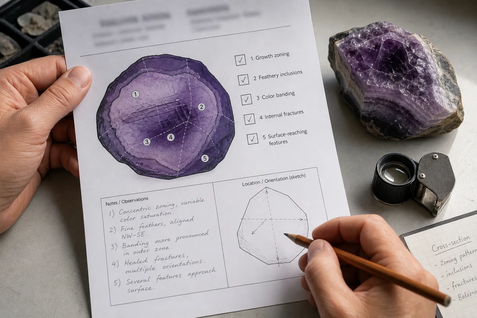
The tempting mistake is to treat an advanced image as an authority object. A three-dimensional map can look decisive because it appears technical, layered, and precise. But a map does not explain itself.
In gemstone conversations, a defect map should not be confused with a certificate, appraisal, or complete authenticity conclusion. It may support an investigation. It may raise better questions. It may show that internal features are unevenly distributed. But without proper source support and method context, it should not be used to assert origin, treatment status, price tier, or buying certainty.
This distinction is especially important for readers who love amethyst both as a mineral and as an object of atmosphere. A specimen can be meaningful in a home without requiring technical proof of every internal feature. It can also be geologically interesting without carrying market claims beyond the evidence.
Think of 3D defect imaging as a lantern inside the stone, not a stamp on the label.
How to Read a Defect Map
If you encounter gemstone defect imaging in an article, lab note, seller explanation, or educational display, read it with a short checklist in mind.
Look for the measured signal.
Does the source say whether the image is based on Raman spectroscopy signals, photoluminescence mapping, another spectroscopic method, or a different imaging approach? If the signal is unnamed, the interpretation should stay modest.
Look for the spatial scale.
Does the map represent a surface, a slice, a volume, or selected points? “3D” should describe how spatial information was collected and reconstructed, not simply decorate the image.
Look for the claim being made.
Showing internal variation is different from establishing authenticity, origin, treatment, or value. The more consequential the claim, the stronger the supporting source needs to be.
Look for comparison context.
A pattern is easier to interpret when the source explains what it is being compared with. Without comparison, the image can remain visually useful while still having limited meaning.
Look for plain limits.
A responsible explanation should say what the map can show and what it cannot settle. If the language jumps from signal mapping to certainty about a stone’s history, slow down.
For collectors, this keeps wonder intact while protecting judgment. The inner life of a crystal can be fascinating without turning every image into a purchase conclusion.
Where This Page Stops
The available material for this page does not include visible, fact-citable references. That creates a firm editorial limit. This article can explain the concept of 3D lattice defect mapping in gemstone-friendly language and describe why spatial distribution may matter. It cannot responsibly make accuracy claims about specific methods, defect-center identification, treatment detection, origin identification, valuation, or lab workflows.
Before this topic is treated as a stricter technical reference, it would need qualified sources from gemological institutes, peer-reviewed spectroscopy or materials-characterization literature, university or museum explainers, standards bodies, or official method documentation. Those sources would need to be used within their proper scope, not stretched into buying certainty or symbolic promises.
The careful answer is this: 3D defect imaging can help reveal the heart of a gemstone by turning hidden internal signal changes into a spatial picture. It can help a reader ask where invisible features are located and how they relate to the stone’s visible character. For an amethyst on a shelf, in a collection, or beside a quiet ritual space, that knowledge belongs to the specimen’s material story; beauty, care, and meaning still require separate kinds of judgment.